One of the critical elements guiding the evolutionary advancement of brains, especially as brains have become super capable in mammals, is how they have acquired increasingly precise function-specific circuitry. As mammalian brains have evolved, so too have the neurons and circuitry responsible for sophisticated motor functions, like grasping a stick, rock, or pen, or playing the piano.
In new research from the lab of Jeffrey Macklis, the Max and Anne Wien Professor of Life Sciences in the Department of Stem Cell and Regenerative Biology, scientists have made an important discovery about those increasingly complicated neurons and brain circuits. The first is that as brains evolved, new subtly different circuits or types of neurons needed to be formed. The second is that to accommodate the new neuron types, mammalian brains ultimately gave the boot to older, less sophisticated, less precise, and less capable neurons. The result: regional evolution of newer types of neurons that will do related functions but with more precision, perfection, and sophistication.
“The axons of these newer types of neurons located in the cerebral cortex of the brain extend remarkably long distances to connect with increasingly specific target neurons in the spinal cord,” explains Macklis. In a new paper published in Cell Reports, Macklis and his team studied mice to discover the evolutionary expansion and neuronal diversification that allows them to perform fine digit manipulation of the forelimb, like picking up a seed. This contrasts with early prototherian mammals like the platypus, where the older neuron types limited motor function such that the forelimbs were capable of more basic cupping movement. The expansion and diversification of this circuitry increased dramatically in non-human primates and humans.
In two earlier papers (Sahni, Cell Reports, 2021a; Sahni, Cell Reports, 2021b), the Macklis lab reported on the basic molecular identification and molecular regulators of the new fancy neurons – the capable, digit manipulation kinds of neurons. They discovered two tiny populations of neurons, split equally between both sides of the mouse brain, totaling just about 4,000 in number out of a total of 85 million neurons in the mouse brain. These neurons are molecularly distinct neuronal subpopulations that innervate the brainstem and cervical cord for more precise, enhanced precision of facial, whisker, and forelimb movement.
Neuronal Crosstalk Keeps the Balance
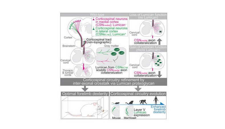
In this new paper, Macklis and co-first authors Yasuhiro Itoh and Vibhu Sahni worked out a surprising and novel form of molecular difference of those highly specialized corticospinal neurons that control voluntary fine motor movement of the face and forelimbs. The roughly 2,000 specialized neurons on each side of the mouse brain secrete the protein lumican at their terminals in the spinal cord which controls the balance of competing connections– “collaterals”– of old and new neuron types, allowing incorporation of newer, more specialized neurons into pre-existing neural circuitry. Lumican helps kick out the evolutionarily older similar function neurons that are not as precise and replaces them with more evolutionarily advanced neurons.
“What’s special about lumican is that it is made several days after the animal’s birth– just when what we conceive of as competition for making connections is occurring, about two weeks after the birth of these specialized neurons,” Macklis explains. It enables crosstalk between these older and newer neuron subpopulations that control mouse corticospinal circuitry refinement and forelimb dexterity. “Lumican essentially biases the competition between the new and old neurons leaving the circuitry,” he adds.
Lumican is part of a family of compounds known as keratan sulfate proteoglycans which are famous for inhibiting neuron growth. In this way, these compounds are very unwelcome in the setting of spinal cord injury because they don’t allow neurons to extend axons and repair damage.
New Targets for Neuron Regeneration
Macklis believes this neuronal competition is just one example of a much broader mechanism for evolutionarily diversifying functional regions of the brain.
“This paper studied the corticospinal motor system, but we believe it has much wider implications for brain development and organization,” he says. “If there are molecules – like lumican – that keep certain kinds of neurons from growing and forming connections, then one might also think about ways to target lumican and other compounds to stimulate neuron growth for regeneration. It might be that there is a whole group of other molecules out there that we would like in the coming decade to discover for regenerative medicine reasons.” Toward that end, Macklis’ team is actively pursuing the idea that molecules secreted by specific populations of growing neurons at their axon tips, or “growth cones,” might be targets for neuronal regeneration.