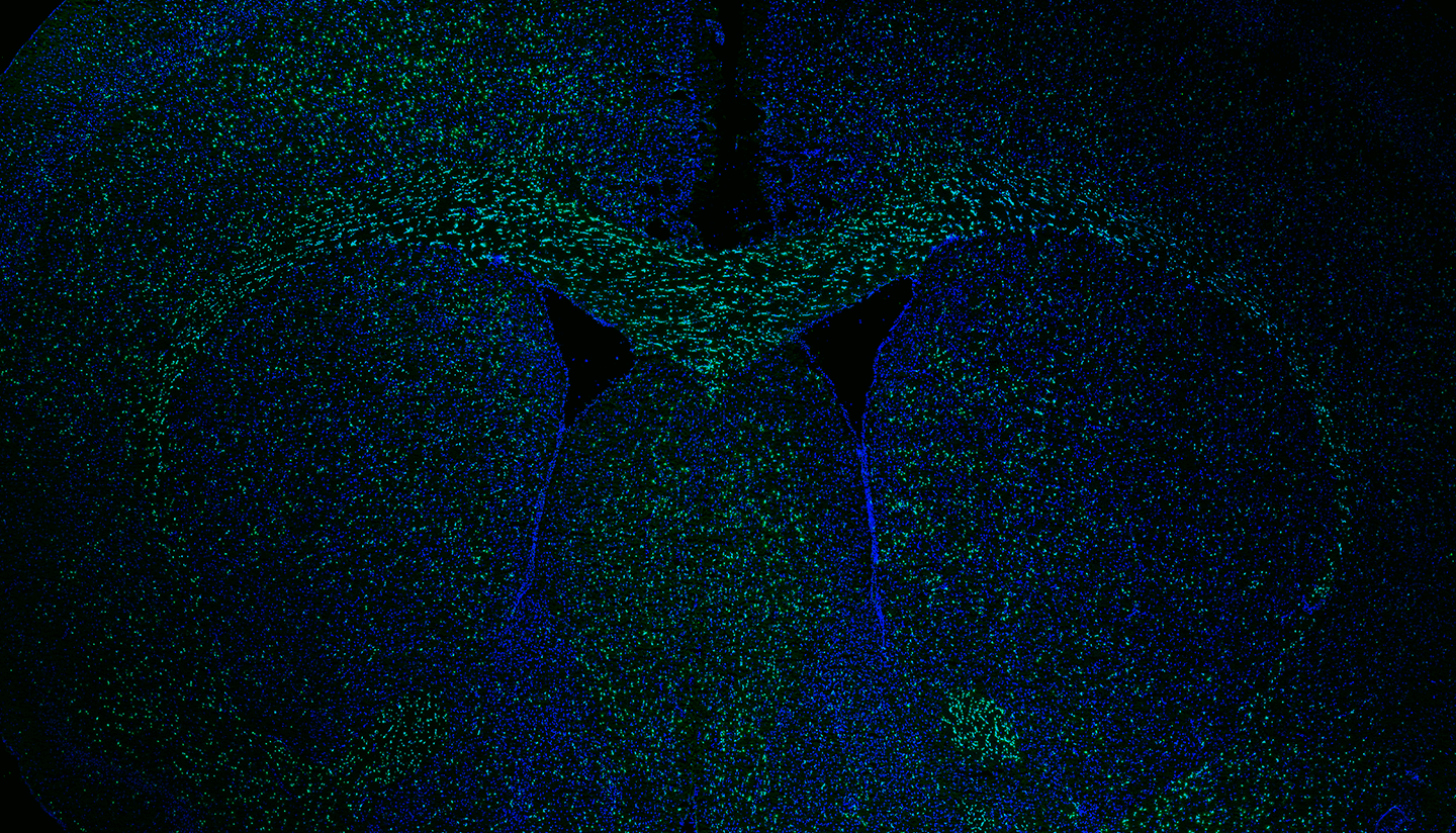
HSCRB researchers have created a highly detailed map of how the mouse brain changes with age. Using single-cell sequencing, the scientists catalogued gene expression in every cell type, providing an unprecedented level of detail on the aging brain. The study, published in Nature Neuroscience, provides a strong foundation for exploring new therapeutic strategies to improve the function of the aging brain.
Over the past decade, researchers have discovered that certain blood factors can regulate the function of the brain, and have a positive effect on the aged nervous system.
“Many studies have asked what’s in blood that alters the brain. But we were also interested in factors in the brain environment itself and how they changed with aging. Interestingly, that had not been thoroughly investigated before,” said senior author Lee Rubin, Ph.D., who is a professor of stem cell and regenerative biology.
What they did
Rubin and his colleagues compared the brains of young and old mice using single-cell RNA sequencing. By analyzing gene expression in tens of thousands of cells, they identified aging patterns in every cell type of the brain, such as the specialized endothelial cells that line blood vessels. The resulting dataset even distinguished among the different sub-types of endothelial cells that line arteries, veins, and capillaries.
Based on the data, the researchers built a theoretical network of how all the different cell types communicate: which molecules are produced by each kind of cell, and which specific cells are targeted by these molecules. Created using powerful computational methods, the communication network identified which interactions change with aging.
What they found
The researchers observed three types of changes in gene expression. For some genes, expression changed in the same way for all cell types. For other genes, expression changed only in specific cell types. Finally, for certain genes, the direction of change was different across cell types.
“Our data show clearly that aging is not a uniform response,” said Methodios Ximerakis, Ph.D., a postdoctoral fellow in the Rubin lab and co-first author of the study. “Each cell type in the brain experiences aging differently as a result of both intrinsic changes and reactions to the changing milieu.”
Why it matters
“One therapeutic approach to treat aging would be to target processes that change in the same way in every cell,” Rubin said. “However, it is also possible to select a particularly important type of cell and try to improve its function. There’s not one approach that would clearly work better than others — each would have particular strengths and weaknesses. This study provides a lot of data to help people decide on the best paths.”
“We now have a clear map of all the genes that change with aging in every single cell type of the brain. Additionally, the intercellular communication network provides a platform that may be useful for identifying novel anti-aging therapeutic targets,” said Ximerakis. “This is a useful resource for those who study aging, and it will hopefully have a big impact on the field.”
Rubin’s therapeutic goal is to improve the cognitive function of the aging brain. Additionally, certain diseases, such as Alzheimer’s disease, have aging as a major risk factor.
“So, our work may also lead to therapeutics to improve brain function in Alzheimer’s disease, as well as other neurodegenerative diseases,” Rubin said.
In this article

Scott Lipnick was a co-first author of the study. The work was supported by Ono Pharmaceutical, the Stanley Center for Psychiatric Research, and the Klarman Cell Observatory.
Source article: Ximerakis, M., Lipnick, S. L., et al. (2019). Single-cell transcriptomic profiling of the aging mouse brain. Nature Neuroscience. https://doi.org/10.1038/s41593-019-0491-3